Nursing Care Plan for Myocardial Infarction

Myocardial infarction (MI)
Myocardial infarction (MI) or acute myocardial infarction (AMI), commonly known as a heart attack, is the interruption of blood supply to a part of the heart, causing heart cells to die. This is most commonly due to occlusion (blockage) of a coronary artery following the rupture of a vulnerable atherosclerotic plaque, which is an unstable collection of lipids (fatty acids) and white blood cells (especially macrophages) in the wall of an artery. The resulting ischemia (restriction in blood supply) and oxygen shortage, if left untreated for a sufficient period of time, can cause damage or death (infarction) of heart muscle tissue (myocardium).
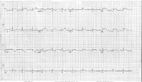
The electrocardiographic result of an acute myocardial infarction is seen below. (See Etiology.)
 The electrocardiogram shows lateral ST-segment elevation that is consistent with a lateral wall acute myocardial infarction.
Myocardial infarction is considered part of a spectrum referred to as acute coronary syndrome (ACS). The ACS continuum representing ongoing myocardial ischemia or injury consists of unstable angina, non–ST-segment elevation myocardial infarction (NSTEMI), and ST-segment elevation myocardial infarction (STEMI). Patients with ischemic discomfort may or may not have ST-segment or T-wave changes denoted on the electrocardiogram (ECG). ST elevations seen on the ECG reflect active and ongoing transmural myocardial injury. Without immediate reperfusion therapy, most persons with STEMI develop Q waves, reflecting a dead zone of myocardium that has undergone irreversible damage and death. Those without ST elevations are diagnosed either with unstable angina or NSTEMI?differentiated by the presence of cardiac enzymes. Both these conditions may or may not have changes on the surface ECG, including ST-segment depression or T-wave morphological changes.
Myocardial infarction may lead to impairment of systolic or diastolic function and to increased predisposition to arrhythmias and other long-term complications.
Assessment
Set basic management to obtain information about the current status of the patient so that all the deviations that occur can be known.
- History or presence of risk factors :
- Arterial disease.
- Previous heart attack.
- Family history of heart disease / heart attack positive.
- High serum cholesterol (above 200 mg / L).
- Smoker
- A diet high in salt and high in fat.
- Obesity. (Ideal body weight = (height -100 ± 10%))
- Women after menopause because estrogen therapy.
- Physical examination: based on cardiovascular assessment may indicate :
Chest pain decreases with rest or administration of nitrate (the most important findings) are often also accompanied by :- Feeling faint and / or death threats
- Diaphoresis.
- Nausea and vomiting sometimes.
- Dispneu.
- Syndrome in various stages of shock (pale, cold, moist or wet skin, lower blood pressure, rapid pulse, decreased peripheral pulse and heart sounds).
- Fever (within 24-48 hours).
- Review of chest pain in relation to :
- Stimulating factor.
- Quality.
- Location.
- Weight.
Painful related to tissue ischaemia secondary to arterial blockage coroner. Possible evidenced by: chest pain with or without spread, face grimacing, restlessness, delirium changes in pulse and blood pressure.
Nursing Intervention
Objectives : Pain decreased after treatment action during ...
Criteria : Chest pain scale decreased for example from 3 to 2, or from 2 to 1, facial expression relaxed / calm, not tense, not restless pulse 60-100 x / minute, blood pressure 120/80 mmHg
Intervention :
- Observation of the characteristics, location, time, and travel is chest pain.
- Instruct the client to stop activity and rest during an attack.
- Help the client to do relaxation techniques, eg deep breathing, distraction behavior, visualization, or the guidance of imagination.
- Keep Olsigenasi with bikanul example (2-4 L / min)
- Monitor vital signs (pulse and blood pressure) every two hours.
- Collaboration with the health team in providing analgesic.
Label: Nursing Care Plan, Nursing Care Plan for Myocardial Infarction
Tidak ada komentar:
Posting Komentar